Abacus dx Cancer Diagnostics – Breast Cancer
Products are for professional/laboratory use only. Breast cancer is the most common cancer affecting women in Australia and New Zealand1 and the second most commonly diagnosed cancer in Australia2. The estimated number of new cases of breast cancer diagnosed in 2019 for Australia was 19,535 and 3,504 for New Zealand1. The number of women diagnosed with breast cancer is expected to increase each year due to the ageing population1. The Cell Marque™ IHC Antibody portfolio offers many of the most valuable, consistent breast markers available in tissue testing.
References:
1. www.breastcancertrials.org.au
2. www.breast-cancer.canceraustralia.gov.au/statistics
Cell Marque™ Immunohistochemistry markers
GATA3 (L50-823)- GATA binding protein 3 or GATA3, is a zinc finger transcription factor and plays an important role in promoting and directing cell proliferation, development, and differentiation in many tissues and cell types. Anti-GATA3 can also be useful in the identification of unknown primary carcinoma when carcinomas of the breast or bladder are a possibility.
Ki-67 (SP6)- The Ki-67 antigen is a nuclear, non-histone protein that is present in proliferating cells. In general, Ki-67 is a good marker of proliferating cell populations. Anti-Ki-67 labeling index has been shown to be a good marker to grade neoplasms including: colon carcinoma, breast carcinoma, and lymphoma
E-cadherin (EP700Y) p120 Catenin (MRQ-5)- E-cadherin is an adhesion protein that is expressed in cells of epithelial lineage. It can be used for the differentiation of ductal (which is membrane staining) vs. lobular cancer of the breast (which is cytoplasmic staining). A combination of E-cadherin and p120 catenin helps distinguish ductal carcinoma of the breast from lobular carcinoma.
HistoCyte breast cancer controls
We are pleased to offer you the HistoCyte product range to add a higher level of trust and ease for your breast cancer testing. Antibodies like ER, PR, and HER2 can require stringent grading standards, and sometimes ideal control tissue is difficult to find- especially in the case of HER2 with 2+ expression.
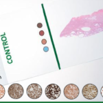
The Dynamic Range blocks from HistoCyte show the full range of expression, including a negative osteosarcoma control. They can be used with IHC and ISH. These are actually cell blocks that were expertly developed to maintain the morphological characteristics of tissue.
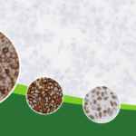
Below is an example of of HER2 antibody stained with IHC and ISH on the HistoCyte HER2 dynamic range block.
ZytoVision ERBB2 Probe for use in breast cancer prognosis
The proto-oncogene ERBB2 is amplified in approximately 20% of all breast cancers and is correlated with a poor prognosis1. Breast cancer patients harbouring ERBB2 amplification are addressed for a targeted therapy with trastuzumab (Herceptin®), a humanized monoclonal antibody directed against the extracellular portion of the ERBB2 protein. This treatment is associated with significantly prolonged overall survival and time of tumor progression2.
The ZytoLight® SPEC ERBB2/CEN 17 Dual Color Probe is designed for the detection of ERBB2 (a.k.a HER2) gene amplification frequently observed in solid malignant neoplasms e.g. breast cancer samples.
References:
1. Brown-Glabermann U, et al. (2014) Oncology (Williston Park) 28: 281-9.
2. Bourdeanu L & Luu T (2014) J Adv Pract Oncol 5: 246-60.


