Cell Marque Peripherin (8G2) Mouse Monoclonal Antibody
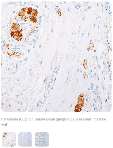
Peripherin is a type III intermediate filament protein localised in the cytoplasm of ganglion cells and in a granular pattern in nerve fibers comprising the peripheral nervous system, which extends to many tissues throughout the body including the salivary gland, small intestine, prostate, stomach, and colon.1
The specific expression of peripherin in ganglia provides utility in the identification of immature ganglion cells in infant or newborn biopsies where there may be morphological ambiguity with other cell types such as endothelial cells, fibroblasts, or inflammatory cells. Likewise, peripherin labeling can be used for visualizing ganglion cell distribution where reduction or loss of observed labeling is correlative with the reduction or loss of ganglion cell presence. The use of anti-peripherin in tracking the reduction or loss of ganglion cells in the submucosal and myenteric layers of the colon wall can act as a valuable tool in identifying patients suspected of recto-sigmoid Hirschsprung disease and other forms of colonic aganglionosis.2-4
References
- Portier, MM et al. Peripherin, a new member of the intermediate filament protein family. Dev Neurosci. 1984; 6:335-344.
- Chisholm, MK and Longacre, TA. Utility of peripherin versus MAP-2 and calretinin in the evaluation of Hirschsprung disease. Appl Immunohistochem Mol Morphol. 2016; 24(9):627-632.
- Holland, SK et al. Utilization of peripherin and S-100 immunohistochemistry in the diagnosis of Hirschsprung disease. Modern Pathology. 2010; 23:1173-1179.
- Solari, V et al. Histopathological differences between recto-sigmoid Hirschsprung’s disease and total colonic aganglionosis. Pediatr Surg Int. 2003; 19:349-354.
THESE PRODUCTS ARE NOT AVAILABLE FOR PURCHASE BY THE GENERAL PUBLIC.


